Long Bone Diagram Inside - Skeletal System Labeled Diagrams Of The Human Skeleton - A typical long bone shows the gross anatomical characteristics of bone.
byAdmin-
0
Long Bone Diagram Inside - Skeletal System Labeled Diagrams Of The Human Skeleton - A typical long bone shows the gross anatomical characteristics of bone.. The long bones are those that are longer than they are wide. Long bones have a thick outside layer of compact bone and an inner medullary cavity containing bone marrow. Long bone anatomy 12 photos of the long bone anatomy long bone anatomy diagram, long bone anatomy worksheet, long bone parts and definitions, long bone structure metaphysis, pediatric long bone anatomy, bone, long bone anatomy diagram, long bone anatomy worksheet, long bone parts and definitions, long bone structure. 4 looking at the inside of the bone.the smallest bone in the human body is called the stirrup bone, located deep inside the ear. A long bone has two parts:
Others are thin, flat, and wide, like your shoulder blades. Long bones are found in the arms (humerus, ulna, radius) and legs (femur, tibia, fibula), as well as in the fingers (metacarpals, phalanges) and toes (metatarsals, phalanges). Related posts of labelled diagram of long bone long bone anatomy. 4 looking at the inside of the bone. Long bone anatomy 12 photos of the long bone anatomy long bone anatomy diagram, long bone anatomy worksheet, long bone parts and definitions, long bone structure metaphysis, pediatric long bone anatomy, bone, long bone anatomy diagram, long bone anatomy worksheet, long bone parts and definitions, long bone structure.
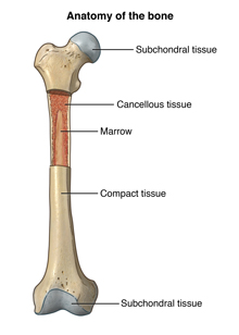
Sinusoidal Vessels And Arterioles In The Bone Marrow A Schematic Download Scientific Diagram from www.researchgate.net Long bones have a thick outside layer of compact bone and an inner medullary cavity containing bone marrow. The interior part of a long bone consists of the medullary cavity. The diaphysis is the tubular shaft that runs between the proximal and distal ends of the bone. The smallest bone in the human body is called the stirrup bone, located deep inside the ear. Add to favorites 0 favs. Important bones diagram human bone anatomy names diagram this arm bones diagram shows all human skeleton diagram of legs human legs bone structure anatomy chart bone anatomy diagram vector illustration of diagram of human bone anatomy royalty free. There is a printable worksheet available for download here so you can take the quiz with pen and paper. Long bones, especially the femur and tibia, are subjected to most of the load during daily activities and they are crucial for skeletal mobility.
Others are thin, flat, and wide, like your shoulder blades.
Diagram of of a long bone. Long bone tissue diagram / introduction to bone boundless anatomy and physiology / a long bone has two main regions:.this is covered by a membrane of connective tissue called the periosteum.beneath the cortical bone layer is a layer of spongy cancellous bone.inside this is the medullary cavity which has an inner core of bone marrow, it contains nutrients and help in formation of cells, made up. Long bone diagram labled : The ends of a long bone contain spongy bone and an epiphyseal line. Related posts of labelled diagram of long bone long bone anatomy. Inside this is a layer of spongy (cancellous) bone which contains red bone marrow. The structure of a long bone allows for the best visualization of all of the parts of a bone (figure 1). Long bone anatomy 12 photos of the long bone anatomy long bone anatomy diagram, long bone anatomy worksheet, long bone parts and definitions, long bone structure metaphysis, pediatric long bone anatomy, bone, long bone anatomy diagram, long bone anatomy worksheet, long bone parts and definitions, long bone structure. Long bone diagram inside : A typical long bone shows the gross anatomical characteristics of bone. The smallest bone in the human body is called the stirrup bone, located deep inside the ear. The femur, or thighbone, is the longest and largest bone in the human body. The structure of a long bone allows for the best visualization of all of the parts of a bone ().
Terms in this set (12). Just in case you get tired of looking at the screen we've provided images and pdf files that you can. Click on the tags below to find other quizzes on the same subject. A long bone has two parts: Inside the diaphysis is the medullary cavity, which is filled with yellow bone marrow in an adult.
Anatomy Of The Bone Johns Hopkins Medicine from www.hopkinsmedicine.org Long bones have a thick outside layer of compact bone and an inner medullary cavity containing bone marrow. The diaphysis and the epiphysis. The diaphysis and the epiphysis. Diagram of of a long bone. Some bones are long and thick, like your thigh bones. The diaphysis is the tubular shaft that runs between the proximal and distal ends of the bone. Inside the diaphysis is the medullary cavity, which is filled with yellow bone marrow in an adult. Choose from 500 different sets of long bone diagram flashcards on quizlet.
A long bone is one that is cylindrical in shape, being longer than it is wide.keep in mind, however, that the term describes the shape of a bone, not its size.
There are two types of bone marrow: A long bone is one that is cylindrical in shape, being longer than it is wide.keep in mind, however, that the term describes the shape of a bone, not its size. Cliffsnotes study guides are written by real teachers and professors, so no matter what you're studying, cliffsnotes can ease your homework headaches and help you score high on exams. The diaphysis is the hollow, tubular shaft that runs between the proximal and distal ends of the bone. Long bones, especially the femur and tibia, are subjected to most of the load during daily activities and they are crucial for skeletal mobility. The diaphysis is the tubular shaft that runs between the proximal and distal ends of the bone. Its lower end helps create the knee joint. A long bone is one that is cylindrical in shape, being longer than it is wide.keep in mind, however, that the term describes the shape of a bone, not its size. The diaphysis and the epiphysis. Long bone diagram labeled find out more about long bone diagram labeled. The diaphysis and the epiphysis. Related posts of labelled diagram of long bone long bone anatomy. Cavity within the shaft of the long bones filled with bone marrow spongy bone layer of bone tissue having many small spaces and found just inside the layer of compact bone.
The diaphysis is the hollow, tubular shaft that runs between the proximal and distal ends of the bone. The smallest bone in the human body is called labeled diagram of an osteon. Long bones, especially the femur and tibia, are subjected to most of the load during daily activities and they are crucial for skeletal mobility. Inside the diaphysis is the medullary cavity, which is filled with yellow bone marrow in an adult. Click on the tags below to find other quizzes on the same subject.
Introduction To Bone Boundless Anatomy And Physiology from textimgs.s3.amazonaws.com Inside this is a layer of spongy (cancellous) bone which contains red bone marrow. The structure of a long bone allows for the best visualization of all of the parts of a bone ((figure)). Long bone anatomy 12 photos of the long bone anatomy long bone anatomy diagram, long bone anatomy worksheet, long bone parts and definitions, long bone structure metaphysis, pediatric long bone anatomy, bone, long bone anatomy diagram, long bone anatomy worksheet, long bone parts and definitions, long bone structure. The ends of a long bone contain spongy bone and an epiphyseal line. Long bone diagram inside : A long bone has a shaft and 2 ends. Just in case you get tired of looking at the screen we've provided images and pdf files that you can. Humerus bone labeled vector illustration diagram long bone type royalty free.
The smallest bone in the human body is called labeled diagram of an osteon.
A long bone has a shaft and 2 ends. The long bones are those that are longer than they are wide. Inside the diaphysis is the medullary cavity, which is filled with yellow bone marrow in an adult. The diaphysis and the epiphysis.the diaphysis is the tubular shaft that runs between the proximal and distal ends of the bone. The diaphysis and the epiphysis (figure 6.3.1). Long bone diagram labled : Diagram of of a long bone. Long bone diagram inside : • reflects the elongated shape rather than the overall size. Long bones have a thick outside layer of compact bone and an inner medullary cavity containing bone marrow. The diaphysis and the epiphysis. Long bones, especially the femur and tibia, are subjected to most of the load during daily activities and they are crucial for skeletal mobility. Long bone labeling diagram quizlet from o.quizlet.com bones are also very good at repairing themselves.
Long bone diagram inside : long bone diagram. The inside of your bones are filled with a soft tissue called marrow.